Growth Disorders
Rapid Growth Problems
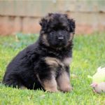
The problems discussed here occur in the younger, rapidly growing German Shepherd. As German Shepherds are far more angulated than most breeds, they can appear to be having serious problems when in many cases they are going through fairly normal stages of development.
Common problems are:
-
Excessive looseness of hocks, can be secondary to excessive depth of hindquarter angulation
or increasing length of hock.
-
Down in pasterns (often seen with 1 above).
-
Flat feet (can be with both 1 and 2).
-
Roached backs – (often associated with 1 above).
-
Lameness – both perception of and real
There is poor general (public) knowledge of growth problems in the GSD. These conditions can include ligament (hocks and pasterns), bone and joint conditions including OCD (elbows, backs), panoestitis, and, less commonly these days, hip dysplasia. Due to the greater angulation of GSD’s compared to other breeds, the perception that there is a problem, particularly during rapid growth, can arise.
Many of these puppies can present as sore with roached backs, very loose in the hocks and/or down in pastern. The age they present can be as young as 12-14 weeks, however, more commonly at around 5-7 months of age. As some of these puppies can appear to be rather loose and or sore, many veterinarians will immediately assume the worst (HD etc) when it can be a relatively easily corrected problem in many cases.
The vast majority of the problems listed above are diet and weight associated, acerbated by (in some cases) the perception of excessive angulation. Most conditions arise following excessive rate of weight gain, usually secondary to the over use of high energy, high density dry foods. Breeders are generally more aware of feeding protocols, and are more likely to keep weights within desirable levels. New owners (ie. the general public) are far more likely to over feed and use expensive high end foods as the more you pay, the better the quality etc...not always so!
Ligament associated problems
Points 1 through 4 are largely associated with ligament issues. Both of these are directly related to rate of weight gain and diet. This is an area that is often poorly understood by owners and veterinarians alike.
Loose ligaments are developed generally from excessive rate of weight gain and, to me, the very high energy dense foods that are over fed. I would remind everyone reading this, that dogs were originally (and largely still are) scavengers – they grew slowly if they could find enough food. German Shepherds are actually very tough dogs and can do well on remarkably little. Add in too rich a food and one pushes the parameters beyond what many puppies can cope with.
Most cases of poor ligamentation that I see are over weight and on high density, high energy puppy foods. Correcting these problems usually involves dropping the diet to an adult diet with a maximum of 22-24% protein and 12-14% protein. The adult foods all have more than enough calcium and vitamin D supplementation to cover growth and have done so for a very long time. Usually the last thing that needs to be added to these puppies diet is calcium - it generally makes matters worse!
However, having mentioned minerals, the vast majority of excessive looseness of ligamentation will respond very well to the addition of zinc, and in some cases iron supplementation. Zinc is a mineral that is often relatively unavailable in the diet, its absorption can be affected by excessive amounts of other minerals, which usually includes calcium as one of the major inhibitors of the uptake of zinc. Zinc is essential for many functions within the body – particularly in enzyme production and ligament strength. Failure to have adequate zinc in the diet can have severe consequences in the developing animals.
Treatment
slow down the rate of weight gain, get the weight back into the accepted normal range. Weight loss at this age is difficult and generally not desired, however, slowing down the rate of gain until they reach the accepted range is the way to go. Do not in general add any calcium, however, the addition of Zinc can be highly beneficial to ligament strength. Ideally the zinc supplemented should be
well absorbed, I suggest using a chelated zinc such as is in Value Plus Organic Iron and Trace minerals supplement. One tablet will cover up to 32 kg daily.
Soreness, usually over the back can be treated with low doses of either Metacam or Carprofen daily for 7-10 days while correcting the diet. If puppies are sore, rest them. Stop them playing with other puppies or adults for about 2 weeks and assess their soreness after this time.
Do not jump into expensive surgeries without checking with your breeder – a mild degree of looseness is expected in younger animals, particularly if they are overweight. The vast majority of puppies with growth associated problems will benefit from rest, correction of diet and/or some mild anti- inflammatory drugs.
Prognosis
Most of these puppies respond very well and visible improvement in both ligamentation and general fitness is usually seen within 2 -3 weeks of correcting diets, rate of weight gain etc. If problems still persist, check with both your breeder and your veterinarian.
Advice to Breeders
breeders should ideally send puppies out of their kennels on a middle range adult dry food. New owners are less likely to cause problems on these foods. High energy, high density puppy dry foods are very easily overfed, particularly by novice owners. A good quality dry food to recommend is the Royal Canin adult GSD dry food (24% protein), or one of the middle of the road adult dry foods. Equally, hand out a copy of the weight chart and remind buyers to look at and use the chart as a guideline.
Rapid growth and bone and joint problems in the young GSD
Osteochondrosis and joint dysplasias have been studied in many species, in particular in pigs. Where the animals were selected for increasingly heavy end weight and rapidity of weight gain, the higher the incidence of symmetrical lesions in certain sites in joints and many growth plates. Experimentally in pigs, the incidence and severity of OCD was directly related to rapid growth ie. rate of weight gain.
When the diet was restricted and grown at a low growth rate, the incidence of OCD was dramatically reduced (almost to zero).
All dog studies in this area have shown to support the concept that the high caloric intake rather than the specific intake of protein, minerals or vitamins influences the frequency and severity of osteochondrosis and HD. The causes of ED while not as thoroughly studied, show similarities and probably similar outcomes.
The common conclusion from studies in dog is that excessive calcium, phosphorus and vitamin D along with a high energy diet and rapid weight gain causing rapid growth, are almost an ideal recipe for pushing the parameters for normal structural growth and joint soundness well beyond their normal limits, resulting in joint disorders. The higher incidence of osteochondrosis in males versus females is probably a direct reflection of this as males are often 1⁄4 heavier than females at any one time, despite being born at a comparative weight.
Equally, this is not to say that genetics does not pay an important part in the body’s structural soundness, however excessive rates of weight gain and thus rapid growth result in pushing the body’s parameters beyond which they can cope, particularly if they were not the most structurally stable to start with. That is, excessive rate of growth and weight will not create severe HD or ED in itself; however, it can make an existing problem considerably worse.
Definitions Dysplasia and OCD overview:
OCD – Osteochondritis Dissecans - genetic, nutritional and rapid growth factors
Osteochondritis (OC) = an error in conversion of cartilage to bone, resulting in thickened and/or necrotic cartilage within joints and growth plates.
Osteochondritis Dissecans (OCD) = progression of OC to cracks, fissures within the cartilage, joint swelling and lameness.
Osteoarthritis (OA) = develops over time if there are remnants of cartilage flaps retained within joints and incongruent joint surfaces developed subsequent to the OCD lesions.
Osteochondritis is a developmental bone disease characterised by defective cartilage in various to multiple joints. The main feature is a failure of the cartilage to properly convert to bone during the growth process. Due to rapid growth, the cartilage cells divide at a normal or increased rate, but the cells do not fully mature and this can result in thickening of the cartilage in various areas of the joint and a delay in bone formation underneath. As the cartilage continues to grow without being normally converted to bone, it looses part of its nutrition because of its increased thickness. Cartilage has no direct blood supply and relies on nutrition from the joint fluid and the underlying bone. The thicker cartilage results in loss of blood supply, in turn causing necrosis (death) of the lower layer of cartilage. Once this happens, the overlying cartilage can separate from the underlying bone (which can be as the result of trauma) causing “flaps” to lift. Such splits allow synovial (joint) fluid into direct contact with underlying bone, creating pain and inflammatory response. This in turn causes the joint capsule to react and become inflamed as well.
Cartilage flaps or loose sections that break away (joint mice) result in continuing pain, and inflammation. Small loose fragments can then attach to the joint capsule or migrate into a connecting tendon sheath causing chronic inflammation around the tendon sheath (more commonly seen in shoulder OCD).
Early lesions of OC can go three ways:-
-
Heal through rest and appropriate treatment such that there is restoration of the ossification process
(ie. normal bone and cartilage development and maturation);
-
Remain unchanged or
-
Progress to OCD, development of arthritis and clinical lameness.
OCD can occur in many different joints, most commonly in the elbows, followed by shoulders, and less commonly in the hocks. The affected joints can be single or bilateral. Different breeds have higher incidences or predilections for various joints to be affected.
Radiographically, cartilage is radiolucent so early changes to cartilage are not readily detectable until there is a delay in the bone formation underneath the cartilage defects, creating a “flattening affect or there are readily apparent defects in the cartilage. Osteoarthritis – develops further over time.
Dysplasia – the term technically means poor or abnormal formation.
“Joint dysplasia might be considered to be the result of an imbalance between weight bearing forces and the maturation of cartilage to bone. If cartilage can convert to bone forming normal shaped articular (joint) surfaces prior to abnormal stresses being placed on the joint, dysplasia can be avoided. However, if the cartilage model becomes malformed prior to the final conversion to bone, the joint will be dysplastic. The severity of each form of dysplasia varies, creating disease with differing clinical significance.” (JP Morgan et al 1999).
Hip Dysplasia
This is a disease that is very common throughout the dog breeds from Cocker Spaniels to Saint Bernards. It is most commonly seen in the heavier bone to muscle ratio breeds where the overall ligamentation is slightly loose.
Hip dysplasia is by definition an ill fitting hip. The hip is a ball and socket joint, and the deeper the socket (ideally sufficiently deep to hold 2/3rds of the head of the femur), the better fit of the femoral head and angle of the neck and the tighter the ligaments, the better the hip.
The various components that combine to give an unstable hip are combinations of the following:-
-
shallow hip socket (the acetabulum),
-
an ill fitting head of the femur (head too small, neck too short and steep)
-
excessive looseness of ligamentation.
Where the socket is very shallow, the ligaments very loose and the femoral head either very steep in the neck or the head very small, these combination of factors lead to instability of the joint. Around the edges of the joint is attached the joint capsule, which in turn is attached to the periosteum. When the edges of the joint capsule are constantly being pulled, the periosteum is lifted and new bone is laid down in an attempt to stabilise the joint.
Rate of Weight Gain
The causes of the development of hip dysplasia, as discussed above, are from a combination of genetic and environmental factors. Rapid weight gain and rate of growth through excessive nutritional intake may cause a disparity of development of supporting tissues. Factors affecting cartilage integrity (thickness and stability) and joint fluid composition, such as repeated trauma from excessive looseness of the joint and /or bacterial infections, can increase joint fluid production, thickening of the joint capsule, resulting in both joint pain and reduction in joint stability. These factors contribute to the development of joint looseness and subsequent subluxation, resulting in early clinical signs and joint changes. Control of the rate of weight gain, while it will not prevent hip dysplasia, it will allow a steady growth pattern allowing the hip structure to mature in concert with the strength of ligamentation in order to minimise excessive stress being placed on the hip joint.
Conversely to osteochondrosis, females in any breed with HD generally have a higher average than males; due it is thought to the influence of female’s hormones. (a 4 point difference on average in the GSD).
Elbow Dysplasia
Results from an incongruity of the joint, mainly as a result of an under development of the ulnar trochlear notch (the inside surface of the joint at the top of the ulna). If the incongruity is minor, there may be minimal changes with no obvious clinical problems. The incongruity results in a step down effect from a poorly developed ulna notch in relation to the weight bearing surface of the top of the radius. If the incongruity is large, a combination of the following 3 major conditions may be seen – UAP, OCD, FCP.
Another form of elbow dysplasia can be seen from radial over growth (relative to the ulna) due to early closure of the distal growth plate of the ulna. Again this is where the cartilage core in this growth plate fails to get adequate nutrition, there is a delay in growth, resulting in a shortened ulna and overgrowth of the radius. This causes incongruity in the elbow and in severe cases may result in bowing of the front legs, lateral luxation of the proximal end (near the elbow) of the radius. The resulting incongruity can again lead to a combination of the above 3 conditions.
FCP (medial Fragmented Coronoid Process)- occasionally, after the anconeal process has united, there can be later development of joint incongrutity can the mico-movement and stresses placed within the joint can result in a fragmented coronoid process – nearly always the medial process.
There are 4 main subgroups of elbow dysplasia – which may occur alone or in combination, generally they are considered inherited in most breeds and the incidence may be adversely affected by incorrect diets in some breeds.
Excessive rate of weight gain can affect the incidence of many elbow conditions, often leading to joint incongruity (uneven rate of growth between the radius and ulnar), and OCD seen particularly in males. (i)UAP – ununited anconeal process – this process is a large triangular shaped piece of bone situated at the back of the elbow joint. This has a separate ossification centre in a handful of breeds mainly larger, heavier breeds - notably the GSD, Labrador, Great Dane, Irish Wolfhound, Newfoundlands, Bloodhound, Basset, Dogge de Bordeaux.This process normally is fully ossified (sealed) by 16-18 weeks of age. This condition is considered inherited with a possible 3 different genes being involved.
The anconeal process forms the hinge at the back of the elbow. When the anconeal process has a separate ossification (growth area) such as in the GSD, it makes this area a target for poor conversion of cartilage to bone in breeds. The combination of the separate ossification centre and a too small circumference of the ulna trochlear notch(inside edge of the back of the joint) results in small movements along the cartilagenous bridge between the ossification centre and the result of the ulna. This results in failure of the anconeal process to unite.
Chronic movement of the UAP results in ongoing development of joint arthritis and further wear and tear of the cartilage surfaces of the joint.
Closure of the ossification centre -The anconeal process in the GSD should be closed by 16-18 weeks of age, definitely by 20 weeks of age. If screening for this X rays taken at 20 weeks should identify a problem, if present.
Clinically signs are seen from as young as 5-6 months with often an intermittent lameness, which is exaggerated by exercise. Full flexion and extension of the elbow will elicit pain. There is often an accompanying lateral bursitis (fluid swelling). X rays of the elbow in the flexed lateral position will readily show whether the process has unified or not. Long standing cases often have boney arthritic changes as well.
Treatment – either a) removal of the process – if done while young, reasonable prognosis with some arthritis later, b) fixation of the loose fragment by a lag screw – reasonable prognosis in early cases. As the anconeal process is not a weight bearing surface, results following early surgery gives much better long term prospects than in the FCP cases (as loss of that process affects the weight bearing surfaces of the joint).
The majority of UAP cases in the GSD have good congruity of the head of the radius relative to the ulnar. Where there is poor congruity, ie. the radius is too long for the ulnar, osteotomy (cutting) of the ulnar to allow it to lengthen naturally is another option.
Affected dogs should not be breed with. UAP is generally considered to be inherited as a separate condition to OCD and FCP in the GSD.
ii. OCD – osteochondritis dessicans (see above as well) – this occurs in many larger breeds, almost any breed greater than 18-20kg at adulthood. There is a higher incidence in males versus females. This can affect many joints, the commonest being the elbow. Breeds that see with a reasonably high incidence of elbow OCD would cover the following :- Rottweiler (high incidence), GSD, Golden Retriever, St Bernard, Great Dane, Border Collie, Rhodesian Ridgeback, Labrador.
Symptoms – generally seen as a shifting lameness in the forequarter from around 5-8 months of age, some joint capsule swelling and usually a turning out of the front legs at stance as the inner edges of the elbow are most commonly affected. The cause of the problem is considered to be due to a faulty blood supply to the joint cartilage secondary to very rapid growth.
Diagnosis - On X ray the signs are often quite subtle in mild cases with minor “fluffiness” and/or flattening of the joint surfaces to the more distinct pot holes of larger lesions. Generally diagnosed by X rays of a straight extended and slightly medially rotated view of the elbow.
Treatment - If this condition is mild, treatment with drugs such as Cartrophen which increases the blood supply to cartilages, can be very effective along with complete rest, slowing down of the rate of weight gain, and low doses of anti-inflammatories. Repair and recovery can take up to 6-8 weeks of age depending on the severity of the condition, very heavy puppies may have to be kept reasonably restricted until 9 months of age by which stage all rapid growth has slowed dramatically. Severe cases of OCD are often found in conjunction with a FCP, and may require surgical intervention. Most cases >80% show good responses to medical, dietary and exercise management.
iii. FCP – fragmented coronoid process of the ulnar – this is generally referring to the medial coronoid process, a process that stabilises the medial edge of the joint. Fragmentation of this process means that the inside edge of the elbow is not stable, hence the very typical lateral rotation of the leg away from the pain. Again, the same age group as above. On X ray the process can be seen as separated on a plate with the elbow extended straight out and a second view with slight medial rotation. If these are diagnosed when young, surgical intervention gives reasonable results, in the older dog where there is considerably secondary arthritic changes, medical management with tablets if probably preferred. Regardless of the treatment, the resulting joint incongruity (unevenness) will lead to ongoing osteoarthritis over time.
iv. Joint incongruity – while most forms of elbow dysplasia can by their development result in joint incongruity, here we are looking at where there has been a possible early closure of a distal growth plate in the foreleg resulting in uneven growth of the radius (usually slightly shorter) in relation to the ulnar. The resulting uneven ends of the bones within the joint can cause excessive wear on cartilages and in the worst cases, force the anconeal process distally (ie. create a UAP). Relatively uncommon in the GSD, however severe cases require surgery.
Other forms of elbow “dysplasia” exist, these forms generally involve the lateral displacement of the top of the radius in relation to the ulnar (giving a cabriole effect) but these very rarely affect the GSD. General Treatment of Elbow Dysplasia – as with any painful bone disorder, regardless of the age presented, common treatment is aimed at pain management, sensible diet and weight control and a restricted, suitable exercise regime. Where there are only minor changes in joint surfaces, medical management and conservative treatment with anti-inflammatory agents and rest is generally all that is needed. Those animals with UAP require surgical intervention to minimise future arthritis. Dogs with FCP or those with loose cartilaginous flaps, should in the younger dog be removed in order to minimise future damage to the joint. However due to the incongruity of the joint, there will be ongoing changes regardless. In the older dog with advanced arthritic changes, medical management and conservative exercise regimes is generally the preferred method of treatment.
Diet and Elbow Dysplasia – while diet may not of itself create elbow dysplasia, it can affect the severity OCD seen. Rapid weight gain will push factors such as joint congruity, as well as blood supply to the cartilages within the joints. Reduce the energy levels in the diets, particularly in the confined dogs – ideally change puppies onto an adult maintenance diet. Affected puppies should have their weights at or below the normal for their breed, sex and age.
Rule outs (differential diagnoses) – not all forelimb lameness is due to elbow dysplasia. Panosteitis and simple injuries should be checked for, particularly where there is a very sudden onset of lameness.
Growth Associated Bone problems
Not all lamenesses in young dogs are genetic, nor involve joints:-
Panoestitis
Affects any of the long bones with a shifting lameness pattern, one or more legs may be affected at any one time. Growth associated problem. Triggering factor(s) unknown. Affected breeds - most commonly seen in the Doberman, less commonly in the GSD (in Australia).
Age affected 5-8 months – can be later as well. Males more commonly affected.
Symptoms – shifting lameness in young dogs with no swelling of growth plates. Affects the long bones of any leg with a shifting lameness pattern, usually only one leg affected at any one time. Pain is exhibited when pressure is applied across the shaft of the affected long bone. Most commonly affected bones are the humerus and femur. X-rays show increased intra-medullary density in the affected long bone.
Treatment - responds well to rest, the use of low grade anti-inflammatories and antibiotic therapy – ie. its treated the same way as a mild bone infection. Dogs usually improve within a few days, but continue antibiotic therapy for a minimum of 3-6 weeks.
Diet – ideally, ease off concentrated high protein foods in maintenance level (22-24% protein, 10-14% fat) in order to slow down the rate of weight gain.
Prognosis - Recovery generally excellent with no long term adverse effects. There is no known hereditary component, and affected animals can be used for breeding.
Contact Details
Chicquita BeckettAdelaide, SA, Australia
Phone : 0433980778
Email : [email protected]